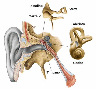
Anatomia del sistema uditivo en
Da "Fisica, onde Musica": un sito web su fisica delle onde e del suono, acustica degli strumenti musicali, scale musicali, armonia e musica.
Jump to navigation Jump to searchHow the ear is structured and how it works
How the ear works is so well known that it does not even seem necessary to explain it. So, to put it simply: the ear is the sensory organ that allows us to hear. Having said that, we now need some more precise information:
- with the term "ear", we do not mean just the visible part (which is called the auricular or pinna) but rather a group of elements that allow us to transform variations in air pressure caused by vibrating sources (a person speaking, a musical instrument, an exploding firecracker, etc.) into electrical impulses that generate sound sensations in the brain;
- technically, we say that the ear is a transducer: i.e. an apparatus that can transport energy from one point in space to another and even transform it into other forms;
- evolutionary mechanisms probably played a role in increasing the efficiency of this transport and creating an ear structure that is generally similar in all mammals.
Let's move on to a description of the anatomy of the various parts of the ear and how they function.
The outer ear
The external ear is made up of the pinna (folds of cartilage) and the meatus (external auditory canal), which is a thin "tube" that ends at the tympanic membrane or tympanum (eardrum). The functions of the pinna are to:
- collect a significant amount of acoustic waves (proportional to the area of the pinna) and lead them through the meatus to the tympanum;
- determine the localisation of a sound source (an operation that could not be carried out with equal precision if we had only one ear instead of two);
- protect the tympanum from mechanical lesions;
- keep the tympanum, which is a very delicate membrane, in a condition of constant temperature, humidity and lubrication in order to preserve its elastic characteristics.
On average, the meatus has a diameter of 7.5 mm and a length of 22 to 25 mm and seems to have no other function except to convey sound waves towards the tympanum. The length of this conduit plays a decisive role in determining the frequency range of maximum auditory sensitivity. With a quick calculation, it is easy to show that, if we treat the conduit as a tube with one open end, it resonates at a frequency of about 2700 Hz (even though, due to the concomitance of other processes, the band of maximum auditory sensitivity is around 3800 Hz).
The middle ear
The middle ear is made up of the tympanum (eardrum), the ossicles (three small bones called the malleus [hammer], incus [anvil] and stapes [stirrup]) and a second membrane, called the oval window, which is the gateway to the inner ear.
- The tympanum is a very thin membrane, held taut by the tympanic muscle, which vibrates when struck by sound waves coming from the outside through the meatus. Thanks to the elastic properties of this membrane, (and an amplification mechanism that we describe below) the tympanum is extraordinarily sensitive. For example:
- a pressure level of 0.2 billionths of atmospheric pressure is sufficient to activate the sound sensation;
- at these pressure levels, the movement of the tympanum is in the order of 10-9 cm (about one tenth of the radius of a hydrogen atom).
- The ossicular chain serves to transfer vibrations from the tympanum to the oval window creating the above-mentioned amplification process of tympanic vibration. One end of the malleus is in direct contact with the tympanum and the other end is "hinged" to the incus, which pushes the stapes against the membrane of the oval window. To be more precise:
- the amplification process is not obtained by the principle of leverage given that the "arm" of the forces acting on the malleus and the incus is about the same (think of how a child sits further away from the fulcrum on a see-saw to balance their weight with the greater weight of their parent). The ossicular chain transfers the almost unaltered force (the gain is about a factor of 1.3) that the tympanum exerts on the malleus directly to the oval window.
- the amplification process is obtained by applying that force to the oval window, the area (about 3.2 mm2 ) of which is about a factor of 17 less than the effective area of the tympanum (i.e. the area of the tympanum that actually vibrates is about 55 mm2). Therefore, the pressure (the ratio between the normal force on the surface and the surface itself) is increased by about a factor of 22 (1.3 for the mechanical effect of leverage times 17 for the effect of area diminution).
- the amplification process of the pressure (and not of the force) is useful because the pressure exerted on the oval window is transferred to the fluid contained in that wonderful structure of the inner ear called the cochlea. Due to their incompressibility, the fluids can, according to Pascal's law, efficiently transfer the pressure exerted on the surface to the all of the fluid within.
- to understand how efficient is the solution developed by nature for transferring sound wave energy to cochlear fluid, consider that if the air were in direct contact with the fluid - due to the bad impedance matching between the two media (air has an acoustic impedance of 41.5 acoustic ohms, while that of cochlear fluid is 143000) - only 1/1000 of the sound wave energy would be transferred to the fluid. The remaining energy would simply be reflected and lost to our hearing!
The inner ear
As said previously, the fundamental structures of the inner ear for the auditory process are:
- The cochlea, which is a spiral-shaped canal carved into the temporal bone. It contains several tunnels filled with endolymphatic fluid (perilymph) that are able to transfer the pressure exerted by the stapes to the oval window, which separates the middle ear from the cochlea.
- The basilar membrane, which is a thin membrane that becomes thicker as it moves away from the oval window and divides the two tunnels (called the scala vestibule and scala tympani) in the cochlea. It terminates with an opening called the helicotrema, which allows the two tunnels to communicate and the perilymph to pass from one part of the basilar membrane to the other.
The function of this membrane is to start oscillating when struck by pressure waves from the cochlear fluid, similarly to a plucked string. The opening of the helicotrema allows pressure waves to propagate both above and below the basilar membrane. The modality of oscillation of this membrane, as a function of the intensity, frequency and spectral composition of sound waves "captured" by the pinnae, plays a fundamental role in the transduction mechanism, which is the basis of perceiving the loudness, pitch and timbre of sounds.
- The organ of Corti, which is a gelatinous structure resting on the basilar membrane and containing cilia directly connected to nerve fibres. The cilia are bent by the oscillatory movement of the basilar membrane. This movement induces the cells of the organ of Corti to generate electrical impulses and send them to nerve endings. These impulses are then conveyed to the brain by the auditory nerve. Therefore, the actual transduction mechanism takes place in the organ of Corti. With regards to this, physiologists have conducted experiments to determine the correlation between the movement of the basilar membrane, the production of nerve impulses and their processing in the brain.
Other functions of the ear
There are two parts of the ear not directly involved in the process of perceiving sound waves. They are:
- the Eustachian tube, which is a conduit that connects the pharynx to the middle ear. It is closed in normal conditions but opens when a pressure difference is created between the two chambers; e.g. when we yawn or swallow. During these moments, air can pass through the middle ear to balance the pressure levels on both sides of the tympanum. This mechanism is very useful when sudden and strong variations of atmospheric pressure occur; e.g. when climbing a mountain or taking off in an airplane (we have all experienced "blocked" ears in these types of conditions). Without this compensation mechanism, the tympanum could be damaged by the pressure difference between its internal and external sides.
- the labyrinth, which is an admirable structure connected to the cochlea and formed by three semicircular, fluid-filled canals oriented in three orthogonal directions. Thanks to the layout of these canals, any movement of the head will set the fluid within the canals into motion with a certain amount of inertia. This, in turn, causes excitation of different neuron populations allowing for the processing of information regarding the state of movement of the head. The central nervous system integrates this information with that coming from our eyes and body muscles to allow us to make voluntary movements and any postural adjustments necessary to maintain our balance. It also allows us to consciously evaluate our position in a geocentric system. It has been noted that in many afflictions of the ear (labyrinthitis, otitis, Ménière's disease), balance is disturbed due to the impaired functioning of the labyrinth.
In-depth study and links
- If you are interested in more in-depth information on the perceptual and physiological aspects of how the ear works, visit the pages on the perception of sound and physiology of the auditory system.
- If you wish to conduct simple experiments on the physiology of the ear, visit this page with Guides to using the Fourier Applet, where you will find applets aimed at highlighting the complexity of the perceptual mechanism.
- If you are interested in other music themes related to consonance and dissonance and issues related to the simultaneous production of many sounds (e.g. in music groups), visit the sections on Critical bands and Masking.
The following is a list of other useful links:
- Localisation of sound sources
- Perception of loudness
- Perception of pitch
- Perception of timbre
- Beats
- Acoustic effects and illusions


